


The brain hemorrhage is a devastating and life threatening catastrophe for a patient and the family. A lot of panic and hopelessness surrounds the family as soon as the diagnosis and the prognosis are revealed to them.
What is Brain Hemorrhage?
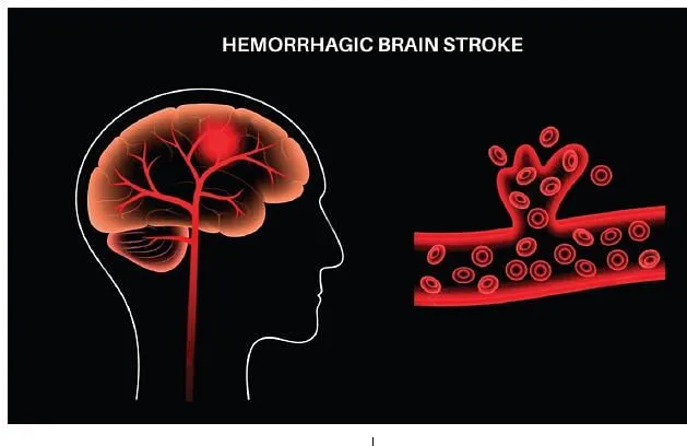
Brain hemorrhage or bleeding means the blood flowing in the lumen of a brain vessel, artery or vein, oozes out due to prick, cut, burst or rupture within or from outside the brain tissue so that the neural tissue is devoid of blood supply and is also pressurized by the weight of the volume of the oozed blood (clot) and the subsequent brain edema (water-logged brain) around the hemorrhagic area.
Let us be aware that the brain hemorrhage or stroke is one of the several types of intracranial (inside-head) hemorrhages which occur by two ways, either insidiously/spontaneously or due to external interference like trauma to head resulting in many different types of intracranial hemorrhages.
But the brain hemorrhage or the hemorrhagic stroke is caused only by the spontaneous/insidious causes like uncontrolled high blood pressure (hypertension) or rupture of a pre-existing aneurysm (segmental arterial saccular or fusiform or blister dilatation) and bleed in an arterio-venous malformation etc.
Physical and Chemical Design of Brain
Though the brain may be an emotional ocean chemically but it has a physical design.
The brain is a semi-solid pinkish-grey dough-like structure, completely occupied by the vascular-arborization, inside the head-bone, cranium. It is enveloped by the three coverings, the outer tougher, middle cobweb but the inner delicate membrane is glued to the brain.
The space, between the outer and the middle coverings, is potential but usually obscured called subdural space. However the space between the middle and the inner coverings is always open and is called the subarachnoid space.
The brain water (cerebrospinal fluid or CSF) is found in the subarachnoid space and it flows over whole brain on all the sides, its base and inside the brain thereby forming water troughs, channels and caves called sulci, fissures, cisterns and ventricles through which run the blood vessels.
These vessels are very different in behaviour than non-brain body vessels in their reaction to the same stimuli like CO2, out-of-lumen blood and O2 causing dilatation and narrowing which increases or reduces the blood supply to the brain that may result in the brain damage called hyperemia, edema, vasospasm, ischemia, infarct etc.
Brain is divided into upper and lower storeyes. The upper one has two hemispheres, each made up of many lobes. The lower brain also has right and left lobes with a central stalk, called brain-stem, connecting it with the upper brain.
The brain lobes consist of brain tissue which comprises billions of pure brain cells (neurons) supported by the supporting brain cells (neuroglia) in the form of hundreds of lobules and nodules (gyrii) separated by the shallow or deep troughs (sulci).
The sulci carry brain water (CSF) through which run the blood vessels just before entering the brain tissue to supply it.
Each brain tissue holds a name for the function it performs like speech area, visual area, hearing area, face area, lip area, thumb area etc. A particular brain area affected by the hemorrhage will affect a particular function which it represents.
Given all the above description, the biological behaviour of the brain appears to be more chemical than physical, that is why the ‘chemical brain’ carries out millions of emotional, cognitive, abstract and physical functions every second in the availability of thousands of neurotransmitters (chemicals).
Origin and types of Brain Hemorrhages
These brain hemorrhages originate spontaneously, internally and insidiously, without any external force, due to rupture of a diseased artery or an abnormal vein outside or inside a brain tissue.
There are three types of hemorrhages by the location of rupture like subarachnoid, intra-parenchymal (intra-cerebral) and both may be combined together.
When the bleeding occurs just outside the brain tissue, that is inside the sulcus, within the brain water –the subarachnoid space or a cistern- before the vessel enters the brain tissue, the bleeding is called spontaneous subarachnoid hemorrhage (SAH).
The causes for this type of brain hemorrhage are the rupture of an aneurysm, bleed in an arterio-venous malformation (AVM), rupture of a diseased artery likely angiopathic (atherosclerosis -unlikely) due to hypertension (high blood pressure >160/90mmHg) and many times a cause may be unknown.
When the bleeding originates within the brain tissue itself, it is known as intracerebral hemorrhage (ICH). The cause of this hemorrhage is primarily uncontrolled hypertension (high blood pressure) and also diseased vessels, angiopathy, within the brain tissue.
Sometimes the bleeding due to rupture of an aneurysm or an AVM may solely occur inside the brain tissue as an ICH. Many times the SAH may extend itself into the brain tissue due to volume-pressure dynamics to form a combination of SAH and ICH together or vice versa can also occur by extension of an ICH into ventricles and subarachnoid space.
However which vessel, normal or diseased, and at what level of high blood pressure it ruptures is still unknown. The blood pressure recorded in the first instance, in the hospital or outside, after the symptoms of brain hemorrhage appear, is not the true blood pressure at which the hemorrhage occurred but this recording is only the blood pressure reading that appeared due to the reaction of the cerebro-cardio-vascular axis after the hemorrhage.
Locations of Brain Hemorrhages
The subarachnoid hemorrhage (SAH) quickly becomes widespread all over-and-under the brain, away from its origin, through the brain water (CSF) to settle in cisterns, fissures, sulci and ventricles as semi-solid clots. Since the bleed is from the vessels anywhere along their course in subarachnoid space, it usually spreads to the whole brain along the brain water.
However, occasionally SAH may become ICH at the site of origin due to aneurysmal direction, burrowing and adherence to the brain. The patient immediately loses consciousness and undergoes even heart (cardiac)-arrest only to revert back spontaneously in most of the cases.
The hemorrhage is picked up by the plain CT scan as a diffuse spread of bleed at the base and side bays of the brain. While as the location of an intracerebral hemorrhage (ICH) is restricted to the site of origin of the bleed only, that is brain tissue, but in many cases due to the larger size of the intracerebral hemorrhage (ICH) the pressure of the bled blood-volume gives vent into the subarachnoid space to become SAH or into ventricles to become intra-ventricular type.
The ICH can occur at many sites in the brain e.g., any lobe of cerebrum, cerebellum, basal ganglia, thalamus, internal capsule, pons, medulla, midbrain, intra-ventricular etc. The serious and life threatening complications arise from such hemorrhages like re-bleeding, hydrocephalus and infarcts which lead to the rise in intracranial pressure (ICP) and squeezing and sandwiching the delicate brain.
Brain hemorrhage Causes and Risk factors
The primary and most common causes of brain hemorrhages are the diseases and abnormalities of the vessels of the brain which burst like a balloon to let blood exude out.
The hemorrhage can occur from the rupture of the pre-existing aneurysms and arterio-venous malformations (AVM) which could either be acquired or may exist since birth.
The untreated and uncontrolled high blood pressures over longer periods of time can be a cause of devastating brain hemorrhage. The underlying cause of uncontrolled high blood pressure may be athero-sclerosis of the cardio-vascular system.
The athero-sclerosis can affect even the vessels of the brain and weaken these to cause rupture anytime in future. The amyloid angiopathy, that weakens vessel walls, is another cause of bleeds which increases with aging and prolonged high blood pressure.
The other well known causes of brain hemorrhages are hemophilia, sickle cell anemia, ingestion of blood-thinners and liver diseases. The risk factors for the brain hemorrhages in the Valley of Kashmir could be intake of intense salt-tea, smoking wet tobacco, ingestion of oily foods, lack of exercise, drug addiction, drugs for blood thinning, untreated high blood pressure, cold remedies in winters etc.
Recognition of Brain hemorrhages
Brain hemorrhage may occur in a person while sleeping, dreaming, sitting, standing, walking, talking, playing, working or exercising, where the patient falls/drops to the ground consciously or unconsciously with paralysis or without paralysis.
The brain hemorrhages occur in all ages, genders and everywhere in the World, more or less in the same proportions. The hemorrhage strikes a person like a ‘bolt from blue’ with severe headache like never before. Most patients lose consciousness before manifesting any other symptom and die suddenly due to massive hemorrhages.
In moderate cases the patient might develop severe headache, vomiting, convulsions, stupor, paralysis of face, arms, legs, loss of speech etc. But headache may be the only symptom in many cases.
Natural History and World View about Brain Hemorrhage
Intracerebral hemorrhage (ICH) accounts for 10–15% of all strokes and carries very high morbidity and mortality rates that have not changed over the last 30 years. About 40% patients of ICH die in a month and up to 65% by the end of the year as studied by R Fogelholm, 2005 and Al-Mufti, 2018 – USA.
The spontaneous subarachnoid hemorrhage (SAH) is associated with severe disability and high mortality rates despite a reduction in case fatality rates over the last four decades. Recent studies have reported mortality rates from 25% at pre-hospital to 18% in-hospital.
Mortality also remains high after discharge from hospital, with rates of 40% within 30 days, 50% within 1 year and an elevated long-term mortality [Nieuwkamp et al -2009; Korja et al -2016; Lantigua et al -2015; Lichtman et al -2011; Huhtakangas et al -2011] in USA.
However both ICH and SAHs usually complicate into frequent re-bleeds within the first 2 weeks, intraventricular hemorrhages (IVH), hydrocephalus and/or vasospasm to produce fatal neuro deficits. The current studies show that the SAH carries a fatality rate as high as 65-80% within a month and one-third of these die suddenly within 24 hours without attending hospital.
Kashmir Overview on Brain Hemorrhages
The fist study related to the brain strokes in Kashmir was published by Sushil Razdan et,al from the SKIMS, Soura in an international journal way back in 1989, titled, “Cerebrovascular diseases in rural Kashmir”.
The study says, “Kuthar valley in the Anantnag district of south Kashmir (North‐west India) was surveyed to ascertain the prevalence of completed strokes. In a population of 63,645, the survey was from July to November 1986; 91 cases of completed stroke were detected giving a crude age-specific prevalence of 143/100,000”.
Although the first aneurysmal SAH, operated by the founder neurosurgeon of Kashmir, Prof. M. Afzal Wani in 1982, went unreported, but the detailed study on SAH in the Valley of Kashmir was published as late as 2011 from the SKIMS, Soura in an international journal.
The study titled ‘Subarachnoid hemorrhage in Kashmir: Causes, risk factors, and outcome’ says that, “the prevalence of aneurysmal SAH in ethnic Kashmiris is high owing to ‘typical diet and habit’ related risk factors and possible genetic factors. SAH, with an incidence of 13/100,000 per year, represents 31.02% of all strokes, a mortality of 36.60%, and good recovery of only 14.99% in Kashmir.
The aneurysmal SAH at 54.35% is the commonest cause of mortality and morbidity. About 19.95% SAHs of all causes ended up in moderate disability. The severe disability occurred in 15.87% and vegetative state was found in 12.56% SAHs. A total of 36.60% of all SAHs died.
Treatment of Brain Hemorrhages
The brain hemorrhage is identified by a CT-scan of the brain and the underlying cause may be revealed by the brain-angiography called digital subtraction, MR or CT cerebral angiography.
If the cause of SAH is an aneurysm or AVM rupture, which is true in most of the SAHs, the treatment is either surgical clipping or Endovascular coiling/stenting or flow diversion of aneurysm and excision or embolization of AVM.
The treatment for the ICH is bimodal that is medical as well as surgical. The existing medical therapy is the prevention of hematoma expansion, blood pressure control and anticoagulation reversal which has been rewarded with improved outcomes.
The role of neurosurgery is still unclear due to the results of the STICH trial (Mendelow, 2011) but the field is rapidly evolving. However the large sized ICHs causing fatal brain pressures and shifts still need surgical decompression to relieve the compression as a life-saving procedure.
Although with moderate sized ICHs, the minimally invasive techniques like neuro-navigation, ultrasound and endoscopic assisted evacuation of hematomas can be promising in selected groups.
Conclusion
The overall outcome of the brain hemorrhages (strokes) presently in the World with all the modern advanced technical gadgetry-of-management remains grim with high fatalities, severe disabilities and low good-recoveries.
But we are hopeful for better treatment options in the near future so that human life thrives healthily.
The author is Prof Head Unit, Department of Neurosurgery and Head Surgical Emergency at SKIMS Soura
Disclaimer: The views and opinions expressed in this article are the personal opinions of the author.
The facts, analysis, assumptions and perspective appearing in the article do not reflect the views of GK.